Species that inform this marker: the community behind every function
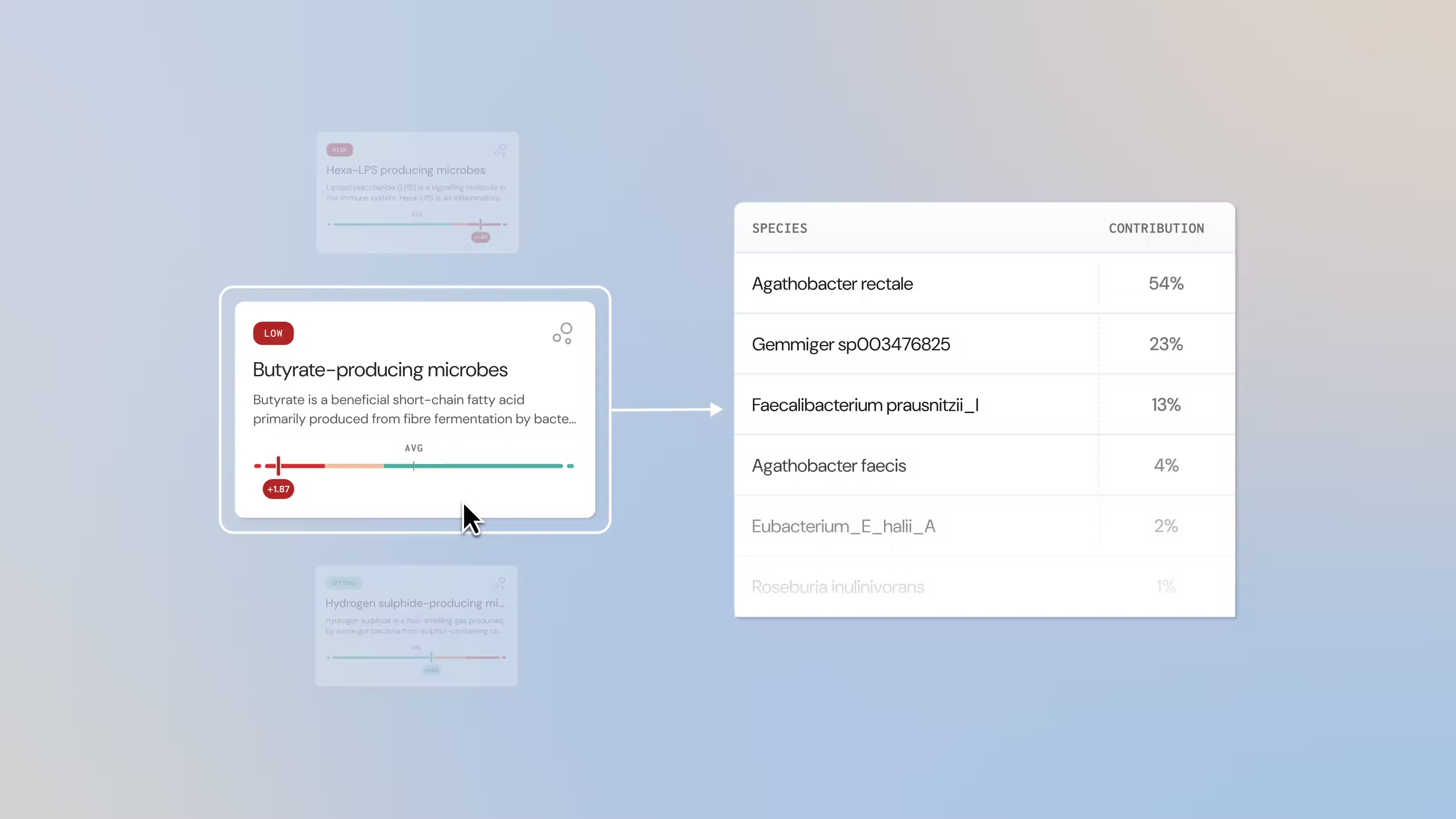
A new section in the Microba microbiome report lists the species contributing to microbiome markers – a practical reminder that the clinical signal is the community’s functional capacity, not a shortlist of well-studied species. For healthcare professionals.
When practitioners first open a metagenomic gut report, the natural instinct is to look for a few familiar, well-studied species and read their presence or absence as the headline. That instinct is a legacy of an earlier era of microbiome science, when lower-resolution methods and a "good versus bad bacteria" narrative encouraged a focus on individual organisms.
The gut microbiome is not a checklist of star species, though – it is a community. Its relevance to health comes less from whether any single organism is present than from what the community as a whole is equipped to do. Reading a report through a handful of representative species is a little like judging a forest by looking for four particular trees.
A new section in the Microba microbiome report is designed to make the whole-community view easy to see. When you open a microbiome marker, you can now scroll to Analytical reasoning, then "Species that inform this marker" – a list of the species contributing to that marker’s function in the patient’s sample. Instead of looking up each organism one by one in Species Explorer, you can see at a glance how many species stand behind a single function.
Why a function is a community, not a shortlist
The reason this matters is a well-established ecological property called functional redundancy – different species often carry the genes for the same metabolic job.
The Human Microbiome Project found that the metagenomic carriage of metabolic pathways stayed stable across healthy people, even though the species present varied widely.1 Later work mapping species to functions showed that roughly half of gut species are metabolic "generalists" rather than specialists.2 A recent analysis of nearly 5,000 gut metagenomes across 28 disease cohorts formally described "functional redundancy clusters" – groups of species that can each perform a given metabolic pathway independently.3
Because the same job is spread across many organisms, two patients with very different species profiles can still have similar functional capacity. That is the point Analytical reasoning makes visible: the capacity behind a marker is shared, and it is rarely the work of one well-known organism.
What the marker measures – potential, not the metabolite
One discipline follows from measuring genes: a metagenomic result describes functional potential – the community’s genetic capacity to perform a function – not a direct measurement of the metabolite. Potential becomes reality only when the right substrates are present, which is why diet is a central lever and why results are interpreted alongside symptoms, clinical history, and diet.
Three functions that show why one species is not enough
Three community functions make the point concrete – two potentially unfavourable, one beneficial. In each, focusing on the well-known species alone can mislead, and Analytical reasoning shows the fuller list of contributors behind the marker.
Hydrogen sulphide – many producers, one easily missed signal
Hydrogen sulphide (H₂S) is a gas made when microbes metabolise sulphur-containing compounds. At physiological levels it may act as an energy substrate for colonocytes and support epithelial signalling. In excess, it may impair colonocyte energy metabolism and compromise the mucus layer and gut barrier.4,5 Crucially, the capacity to make H₂S is spread across many species – for example Desulfovibrio piger, Bilophila wadsworthia, Gordonibacter pamelaeae, Intestinibacter bartlettii, Flavonifractor plautii and Turicibacter sanguinis, among others (the list is not exhaustive; see the marker card guide).
This is exactly where a celebrity-species view can fall short. To illustrate the principle: a test that assesses only Desulfovibrio species could read as "optimal" even when the wider community has a high capacity to produce H₂S. A community-level assessment captures that broader functional potential. "Species that inform this marker" shows which producers are contributing to the H₂S marker, so you can see which organisms make up the H2S-producing community in each sample.
Diet is a meaningful modifier. In a human crossover study, ex vivo faecal H₂S production was higher on an animal-based, low-fibre diet than on a plant-based, high-fibre diet – but with substantial variability between individuals, and 2 of 11 participants produced more on the plant-based diet.6 That variability is a further reason to measure an individual’s community rather than assume how it will behave.
Hexa-acylated LPS – not all endotoxin is equal
Lipopolysaccharide (LPS) shows a subtler trap. LPS is an outer-membrane component of Gram-negative bacteria, but its inflammatory potency depends on the acylation state of its lipid A. Hexa-acylated LPS – the six or higher-chain form – is a strong agonist (activator) of the innate immune receptor TLR4.7 Under-acylated forms with fewer chains are typically weak, and some actively antagonise (inhibit) TLR4. In LPS purified from healthy human faecal samples, it was found that total LPS was largely TLR4-silencing, because much of it is under-acylated LPS from Bacteroidales.8
The functional consequences track this structure. In a controlled study in mice, hexa-acylated LPS from Escherichia coli impaired gut barrier function and worsened glucose control, while an equal dose of an under-acylated, TLR4-antagonising LPS did not – and could even offset the harmful type.9 So counting "Gram-negative bacteria" or "total LPS", or reading a single marker species, tells you little about inflammatory potential. What matters is the community’s capacity to make specifically hexa-acylated LPS.
Producers include Escherichia coli, Klebsiella pneumoniae, Klebsiella variicola, Citrobacter freundii, Pseudomonas aeruginosa and Raoultella ornithinolytica, among other Gammaproteobacteria (list not exhaustive; see the marker card guide). A higher capacity to make hexa-acylated LPS has been associated with inflammatory conditions. In a metagenome-wide analysis of Crohn’s disease and ulcerative colitis, Enterobacteriaceae such as E. coli – and their hexa-acylated LPS-biosynthesis pathways – were overrepresented in patients.10
A large study of inflammatory arthritis (221 patients and 219 controls) implicated community-level pathways, including hexa-acylated LPS biosynthesis, in rheumatoid arthritis and ankylosing spondylitis.11 Assessing capacity across all the organisms that carry the relevant genes – not one representative species – is what makes the result meaningful.
Butyrate – the beneficial mirror image
The same logic applies to functions we want to see. Butyrate is a short-chain fatty acid and a primary energy source for colonocytes. It is associated with gut barrier integrity12 and immune regulation, including the promotion of regulatory T cells and anti-inflammatory signalling.13 The community’s potential to make butyrate reflects the combined capacity of many producers, rather than just a few organisms. It is also often supported by cross-feeding, where one species’ metabolites become another species’ substrate for butyrate production.
Examples of butyrate producers include multiple Faecalibacterium prausnitzii clades, Roseburia hominis, Anaerostipes hadrus, Coprococcus_A comes, Gemmiger formicilis and Odoribacter splanchnicus (list not exhaustive; see the marker card guide). So a microbiome that lacks a celebrated producer such as F. prausnitzii may still have ample community-level butyrate capacity through other species. Conversely, the presence of one well-known producer does not guarantee sufficient capacity. Judging butyrate from one or two named organisms misses this.
About the species names you’ll see
The "Species that inform this marker" list uses names from the Genome Taxonomy Database (GTDB), the reference taxonomy behind the report. Genome-based classification sometimes splits a single named species into multiple species. When it does, GTDB adds a letter suffix – for example Faecalibacterium prausnitzii_C.
Species without a formal name yet are given an alphanumeric placeholder, such as Faecalibacterium MIC7145. These are not typos or strain codes; they are precise, species-level identities. Seeing several closely related names under one function is normal, and it reflects the resolution of shotgun metagenomic sequencing.
What this means for interpretation
The shift is from "Which species are present?" to "What is this community equipped to do, and in what context?" In practice, that means reading functional capacity at the community level using the microbiome markers, and scrolling to Analytical reasoning to see the contributors behind each one.
It means interpreting alongside symptoms, diet, medications and clinical history – because whether that potential is realised depends partly on substrate availability, which makes diet both context and a possible intervention lever. And it means using functional markers to guide personalised, iterative strategies – for example, increasing fibre diversity to support the butyrate-producing community, or moderating sulphur-rich animal protein where H₂S capacity is high – and to track change over time.
The bottom line
The microbiome is a community, and what it is equipped to do can be more informative than the presence of a few individual species. Functional redundancy means many species can contribute to the same function, so different species profiles can result in a similar functional capacity. Single-species or "good/bad" reads can mislead in both directions – H₂S may look "optimal" on one species while being high across the community, and butyrate capacity may be adequate even without a well-known producer.
A molecule’s effect can also depend on its structure; for LPS, the hexa-acylated form is the one most relevant to inflammatory potential. "Species that inform this marker" makes the full set of contributors visible, so interpretation rests on the whole community rather than a shortlist.
References
- Huttenhower C. et al. Structure, function and diversity of the healthy human microbiome. Nature 486, 207–214 (2012). https://doi.org/10.1038/nature11234
- Vieira-Silva S. et al. Species–function relationships shape ecological properties of the human gut microbiome. Nat. Microbiol. 1, 16088 (2016). https://doi.org/10.1038/nmicrobiol.2016.88
- Jiang Y., Che L. & Li S. C. Deciphering the personalized functional redundancy hierarchy in the gut microbiome. Microbiome 14, 17 (2025). https://doi.org/10.1186/s40168-025-02273-w
- Blachier F. et al. Production of hydrogen sulfide by the intestinal microbiota and epithelial cells and consequences for the colonic and rectal mucosa. Am. J. Physiol. Gastrointest. Liver Physiol. 320, G125–G135 (2021). https://doi.org/10.1152/ajpgi.00261.2020
- Ijssennagger N. et al. Gut microbiota facilitates dietary heme-induced epithelial hyperproliferation by opening the mucus barrier in colon. Proc. Natl. Acad. Sci. U.S.A. 112, 10038–10043 (2015). https://doi.org/10.1073/pnas.1507645112
- Teigen L. et al. Differential hydrogen sulfide production by a human cohort in response to animal- and plant-based diet interventions. Clin. Nutr. 41, 1153–1162 (2022). https://doi.org/10.1016/j.clnu.2022.03.028
- Zamyatina A. & Heine H. Lipopolysaccharide recognition in the crossroads of TLR4 and caspase-4/11 mediated inflammatory pathways. Front. Immunol. 11, 585146 (2020). https://doi.org/10.3389/fimmu.2020.585146
- d’Hennezel E., Abubucker S., Murphy L. O. & Cullen T. W. Total lipopolysaccharide from the human gut microbiome silences Toll-like receptor signaling. mSystems 2, e00046-17 (2017). https://doi.org/10.1128/mSystems.00046-17
- Anhé F. F., Barra N. G., Cavallari J. F., Henriksbo B. D. & Schertzer J. D. Metabolic endotoxemia is dictated by the type of lipopolysaccharide. Cell Rep. 36, 109691 (2021). https://doi.org/10.1016/j.celrep.2021.109691
- Khorsand B. et al. Overrepresentation of Enterobacteriaceae and Escherichia coli is the major gut microbiome signature in Crohn’s disease and ulcerative colitis. Front. Cell. Infect. Microbiol. 12, 1015890 (2022). https://doi.org/10.3389/fcimb.2022.1015890
- Thompson K. N. et al. Alterations in the gut microbiome implicate key taxa and metabolic pathways across inflammatory arthritis phenotypes. Sci. Transl. Med. 15, eabn4722 (2023). https://doi.org/10.1126/scitranslmed.abn4722
- Wang R. X., Lee J. S., Campbell E. L. & Colgan S. P. Microbiota-derived butyrate dynamically regulates intestinal homeostasis through regulation of actin-associated protein synaptopodin. Proc. Natl. Acad. Sci. U.S.A. 117, 11648–11657 (2020). https://doi.org/10.1073/pnas.1917597117
- Singh N. et al. Activation of Gpr109a, receptor for niacin and the commensal metabolite butyrate, suppresses colonic inflammation and carcinogenesis. Immunity 40, 128–139 (2014). https://doi.org/10.1016/j.immuni.2013.12.007
- Costabile G. et al. Daily profiles of plasma short-chain fatty acids after the intake of three different cereal fibers: a randomized controlled study. Eur. J. Nutr. 64, 217 (2025). https://doi.org/10.1007/s00394-025-03741-7